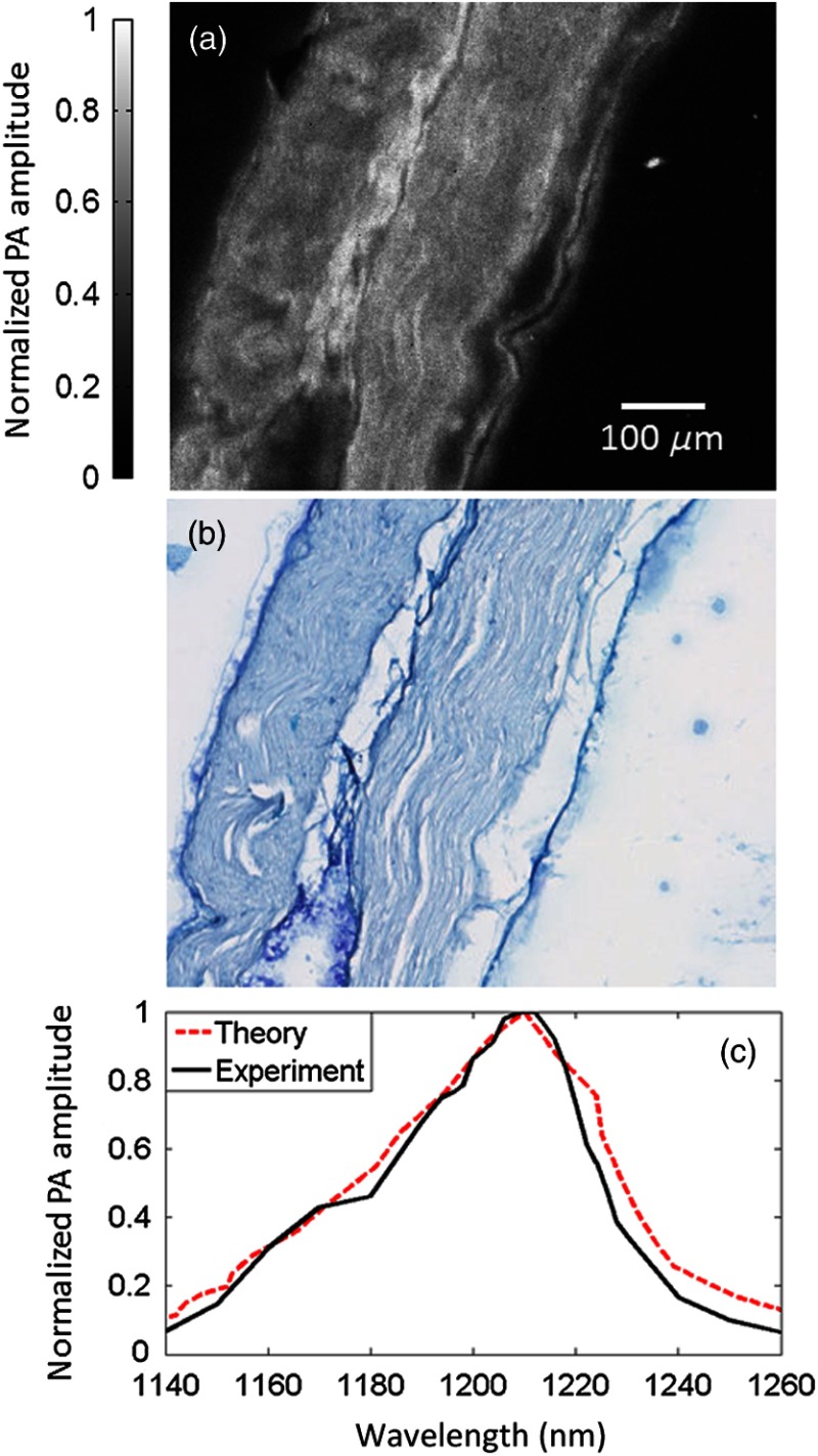
Fig. 4.
(a) Photoacoustic (PA) image of a sectioned, unstained sciatic nerve. (b) Bright-field optical image of a sectioned nerve stained with luxol fast blue, targeting myelin, and cresyl violet, targeting nuclei and Nissl bodies. (c) The PA spectrum of a whole nerve matches the absorption spectrum of lipids.

