
Fig. 4.
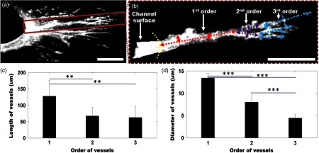
(a) Cross-sectional projection of the sprouting vessel. (b) The highlighted vessel in (a) is shown separately to illustrate the vessel orders and diameter and length measurements. The yellow dashed line marks the surface of the channel seeded with endothelial cells. The first-, second-, and third-order vessels are marked with red, purple, and blue dashed lines, respectively. (c, d) Quantitative comparison of vessel lengths and diameters for different vessel orders. Student’s t-tests were performed for statistically analysis (** and ***). Scale bar is 100 μm.