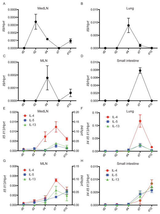
Figure 1. IL-9 expression precedes IL-4, IL-5 and IL-13 during N. brasiliensis infection.
(A–D) C57BL/6 mice were subcutaneously infected with 625 L3 N. brasiliensis larvae. MedLN (A), lung (B), MLN (C) and small intestine (D) were collected and homogenized at different days p.i for assessment of il9 mRNA expression by real time RT-PCR. The experiment was performed two times with similar results with 2–3 mice per day p.i. Statistically significant p values were determined by one-way ANOVA when comparing basal expression (d0) with at least one other time point for each gene.
(E–H) Same samples as above analyzed for Il4, Il5 and Il13 mRNA expression. Data represent the mean +/− SEM ratio of cytokine gene to Hprt expression as determined by the relative quantification method (ΔΔCt). The experiment was performed two times with similar results with 2–3 mice per day p.i. Statistically significant p values were determined by one-way ANOVA when comparing basal expression (d0) with at least one other time point for each gene.

