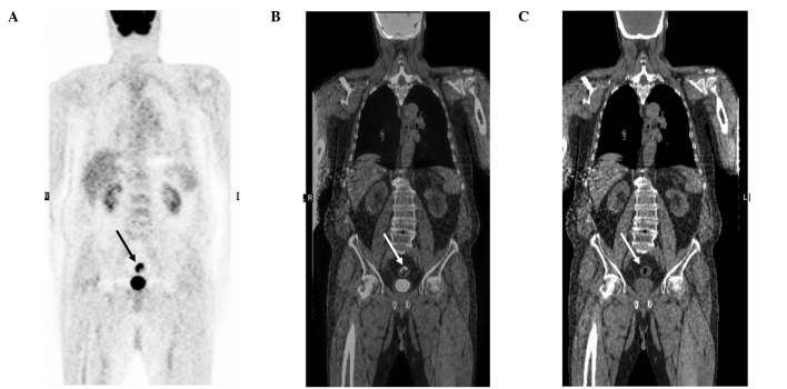
Figure 2.
A 79-year-old male was referred for the assessment of a pulmonary nodule observed on CT. (A) Coronal FDG-PET, (B) PET/CT and (C) CT slices demonstrate intense FDG uptake in the proximal region of the colon (arrows). On colonoscopy, a large ulcer was found in 15 cm into the colon. The final diagnosis was of an adenocarcinoma of the colon. FDG PET/CT, fluorodeoxyglucose positron emission tomography/computed tomography.

