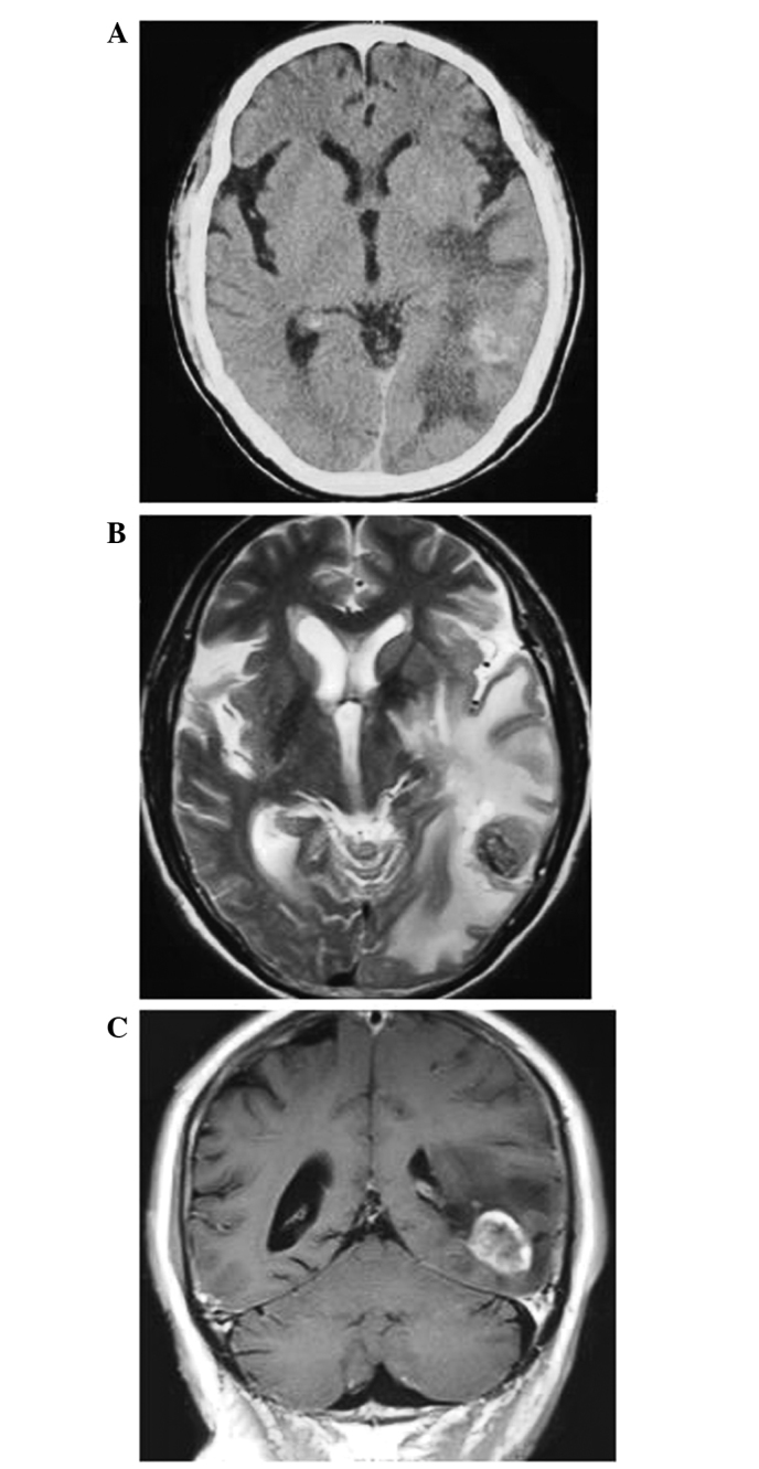
Figure 1.

Prior to treatment, (A) axial CT, (B) axial MRI T2-weighted and (C) coronal MRI T1-weighted scans reveal brain metastasis in the left temporal lobe and cerebral edema located around the lesion. CT, computed tomography; MRI, magnetic resonance imaging.
