
Figure 2.
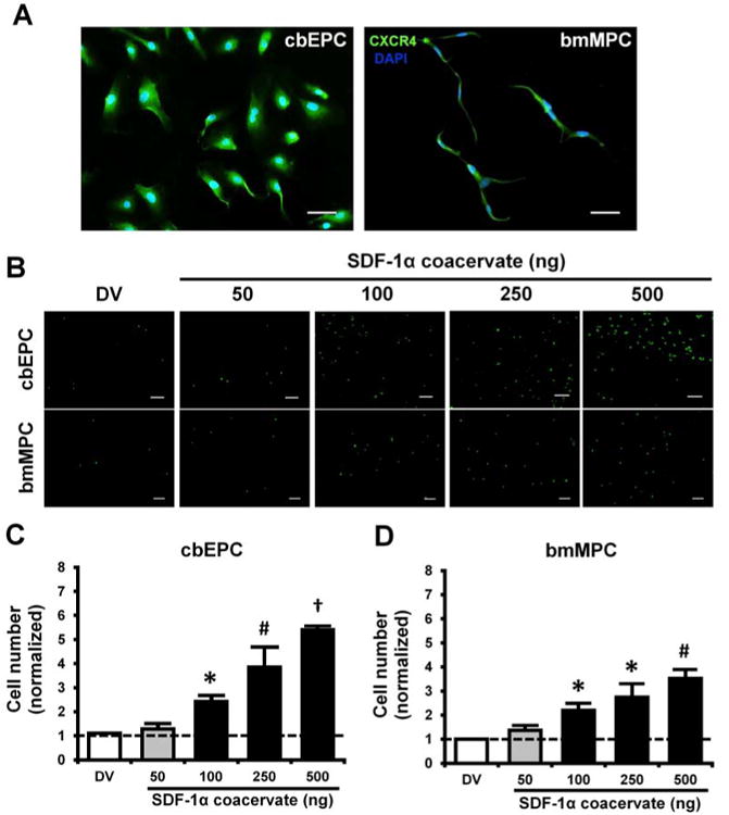
Characterization and chemotaxis of progenitor cells. (A) CXCR4 expression of cbEPCs and bmMPCs, (B) Fluorescent images of migrated progenitor cells on the bottom of transwell insert membrane. Scale bar = 200 μm. (C) and (D) Quantification of migrated cells. Cell numbers in each group were normalized by those in basal medium group. *p < 0.05 (compared to DV and 50), #p < 0.05 (compared to DV, 50, and 100), †p < 0.05 (compared to all other groups) (n = 4).