
Figure 6.
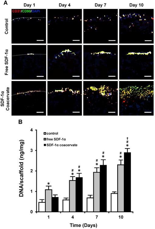
Recruitment of progenitor cells into the SDF-1α coacervate-laden scaffolds. (A) Immunofluorescent staining for CD31 (cbEPC marker, red), CD90 (bmMPC marker, green), and DAPI (nuclei, blue). Dashed lines represent the border between the outer region (top) and the scaffolds (bottom). Scale bar = 100 μm (all). (B) Quantification of the number of cells in scaffolds. The number of cells was normalized by the wet weight of scaffolds. *p < 0.05 (compared to control), #p < 0.05 (compared to day 1), †p < 0.05 (compared to day 4) (n = 4).