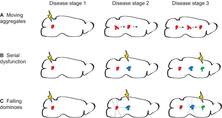
Fig. 4.
Models of protein aggregate spread. Three models of how the localization of aggregates in the brain might change as neurodegenerative disease progresses. (A) Moving aggregates. Aggregates generated in one region physically move to another, covering a large distance. (B) Serial dysfunction. Due to network dysfunction, conditions inducing de novo synthesis of protein aggregates move across the brain. (C) Falling dominoes. Similar to serial dysfunction, where conditions permissive to aggregate formation move, but with the distinction that misfolded protein aggregates seed the formation of new aggregates in close proximity, creating a chain reaction. The lightning bolts represent induction of conditions in different brain regions that accommodate the appearance of protein aggregates. Conditions might include aberrant network activity or impairment of protein quality-control machinery. The arrows in A show that an aggregate formed in the left end of the brain moves towards the right end. The dots in C represent small oligomeric misfolded protein seeds, which spread and can form full size aggregates once permissive conditions are present (lightning bolt) but not in areas without permissive conditions (above and below the aggregates).

