Fig. 3.

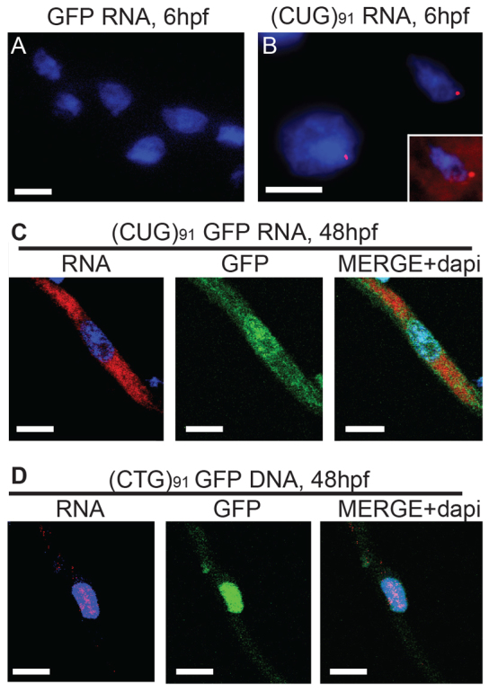
RNA foci formation in GFP(CUG)91 mRNA-injected embryos. (A) In situ hybridization using a Cy5–2-O-methyl (CAG)5 RNA probe in GFP mRNA-injected embryos at 6 hpf. There was significant GFP visible in all the cells (not shown) but no foci. (B) In situ hybridization using a Cy5–2-O-methyl (CAG)5 RNA probe in GFP(CUG)91 mRNA-injected embryos at 6 hpf. Foci were readily visible in many nuclei. In addition, there was significant diffuse nuclear and cytoplasmic Cy5 signal above that seen in GFP mRNA-injected embryos (inset). (C) Dissociated myofibers from GFP(CUG)91 mRNA-injected embryos isolated at 48 hpf. Nuclear RNA foci were rarely seen, but RNA was still present diffusely in the cytoplasm and (to a lesser degree) the nucleus. (D) Dissociated myofibers from GFP(CUG)91 DNA-injected embryos isolated at 48 hpf. Nuclear RNA foci were readily visible in GFP-positive myofibers, with very little visible diffusely in the nucleus or cytoplasm. Scale bars: 20 μm.
