
Figure 5.
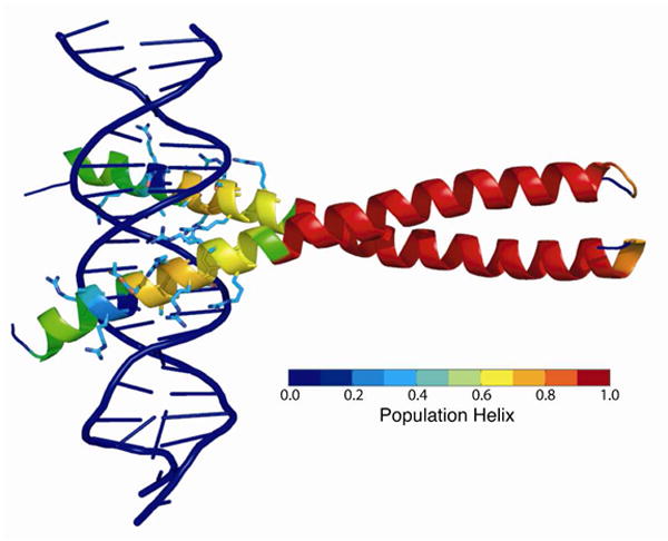
The fractional helicity of each residue observed in trajectory 2, the trajectory in the best agreement with experimental NMR data, is mapped onto the crystal structure of the GCN4 bZip domain bound to DNA (PDB code 1YSA) according to the color bar shown. Side chains are displayed for residues which make contacts with the DNA.