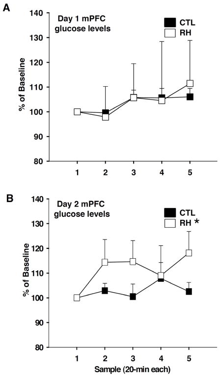
Fig. 2.
Medial prefrontal cortex (mPFC) extracellular fluid (ECF) glucose levels, measured before, during, and after Set-Shift testing of control animals (CTL) and animals with prior history of recurrent hypoglycemia (RH) on day 1 (A) and day 2 (B) of the behavior task. ECF samples were collected in 20 min bins. Data are expressed as percentage of baseline + SEM. Asterisks indicate significantly different group findings (p<0.05).

