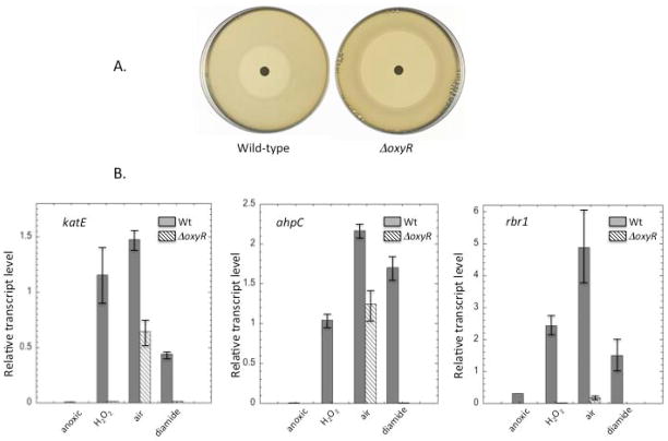
Fig. 8. Role of OxyR in H2O2 stress.
(A) Wild-type and ΔoxyR strains were spread on anoxic BHIS plates and exposed to a filter disk containing H2O2. The zone of inhibition after 24 hours of incubation was 26 +/− 2 mm for wild-type cells and 50 +/− 3 mm for the ΔoxyR strain. (B) The katE, ahpC, and rbr1 genes were induced in an OxyR-dependent manner upon anoxic treatment with H2O2 or diamide. Induction upon aeration had OxyR-dependent and –independent components. Transcripts were measured by qRT-PCR under anoxia, after anoxic treatment with 100 μM H2O2 for 30 min, after aeration for 45 min, or after anoxic treatment with 1 mM diamide for 15 min. Error bars represent SD from the mean of three independent experiments.

