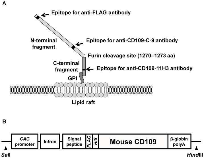
Figure 1. Structure of CD109 cell-surface glycoprotein.
A, Schematic illustration of FLAG-tagged human CD109 (FLAG-hCD109) structure on cytoplasmic membrane. Anti-CD109-C-9 mAb, anti-FLAG mAb and anti-FLAG pAb can detect 180-kDa N-terminal fragments; anti-CD109-11H3 mAb can detect 25-kDa C-terminal fragments. B, Schematic illustration of construction of FLAG-tagged mouse CD109 (FLAG-mCD109) transgene.

