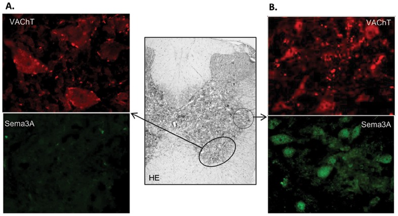
Figure 3. Semaphorin 3A (Sema3A) in the intermedioratelal nucleus of the spinal cord in PVL rats.
(A) Magnifications (400×) of the ventral horn area of the spinal cord (middle gray color image at 4×), containing cholinergic neurons, showing strong VAChT immunofluorescent signal and null immunoreactivity for Sema3A. (B) Magnifications (400×) of the spinal cord intermediolateral nucleus containing cholinergic preganglionic sympathetic neurons. These neurons show positive immunofluorescent signal for both VAChT and Sema3A. HE, hematoxilin-eosin staining.

