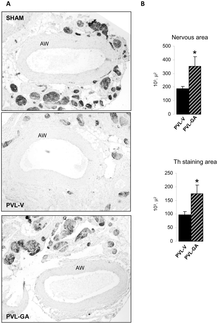
Figure 6. Analysis of sympathetic atrophy in the superior mesenteric artery (SMA) after gambogic amide administration.
(A) Representative images of tyrosine hydroxylase (Th) immunostaining at 40× showing transversal sections of complete arterial wall (AW) surrounded by nervous structures from sham (showed as a normal state reference), PVL vehicle (PVL-V) (n = 6), PVL gambogic amide (PVL-GA) (n = 6), *p<0.05, **p<0.001, compared to PVL-V. (B) Bar diagrams showing immunohistochemical quantitation of total nervous area and Th staining area in nerves surrounding the SMA from experimental groups PVL-V and PVL-GA.

