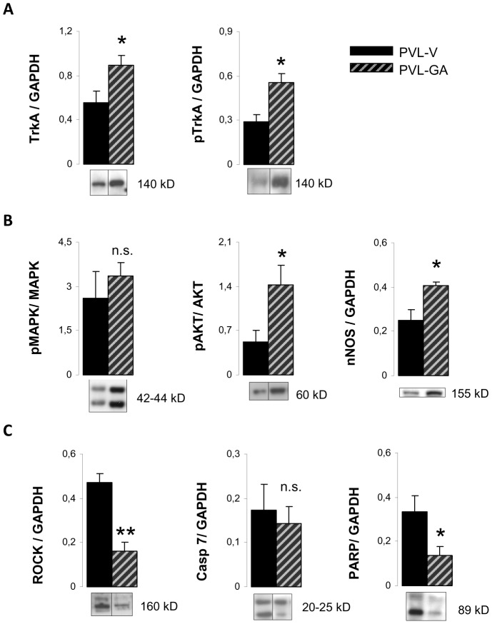
Figure 7. Protein expression in the superior mesenteric ganglion after gambogic amide administration.
Bar diagrams showing quantitation by Western blot analysis of (A) tyrosine kinase receptor A (TrkA), phospho-TrkA (pTrkA), (B) the ratio of phosphorylated and total forms of mitogen activated protein kinase (MAPK) and protein kinase B (AKT) and neuronal nitric oxide synthase (nNOS), (C) Rho kinase (ROCK), cleaved caspase 7 (Casp-7) and poly(ADP-ribose)polymerase (PARP) in PVL treated with vehicle (PVL-V) (n = 8) or gambogic amide (PVL-GA) (n = 8). Representative Western blots are shown below. Only bands corresponding to the phosphorylated form are exemplified in the case of the quantitation of ratios. Grouping of bands from different parts of the same gel are denoted by dividing lines.

