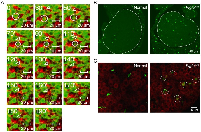
Figure 6. Macrophages involved in clearance of dead oocytes in normal and Figla null ovaries.
(A) Time-lapse recording of a normal Figla-EGFP/Cre; mTomato/mEGFP newborn ovary captured a destruction process of an oocyte (circle). A somatic cell (arrowhead) closely associates with the oocyte and appears to phagocytize the remains of the degrading oocyte. (B) Normal or Figla null newborn ovary sections were immuno-stained with F4/80, a macrophage specific marker. Ovarian tissue is outlined by dotted line. (C) Oocytes and macrophages were stained by Mvh (red) or F4/80 (green), respectively. Macrophage-engulfed degrading oocytes were observed in Figla null (dotted circles), but not normal ovaries.

