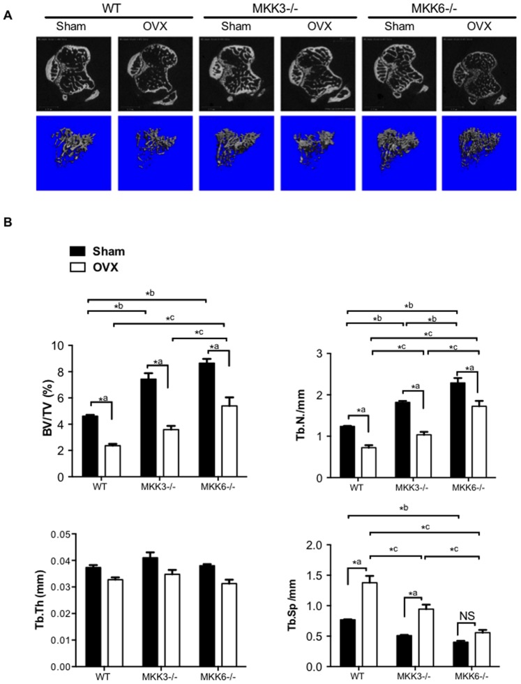
Figure 4. Ovariectomy-induced bone loss in MKK3−/− and MKK6−/− mice.
(A) WT, MKK3−/− and MKK6−/− mice were subjected to ovariectomy and bone loss was calculated using micro-CT analysis (n = 5/group). Representative images are shown. Significant bone loss was observed in all three groups. (B) Bone volume and trabecular number were elevated in MKK3−/− and MKK6−/− sham-treated or OVX mice compared with respective WT controls. Trabecular thickness was similar in sham-treated and OVX mice. Trabecular separation was significantly higher in WT and MKK3−/− mice after OVX. MKK3- and MKK6-deficient mice had high bone mass phenotype compared with WT in sham-treated or OVX groups.

