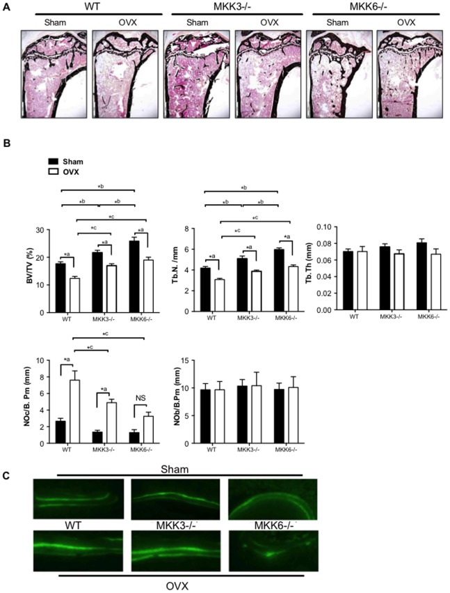
Figure 5. Histomorphometric analysis of bone in ovariectomized MKK3−/− and MKK6−/− mice.
(A) Tibial bone sections from sham and OVX-treated mice were stained and representative figures are shown (n = 5/group) (B) Histomorphometric analysis showed that bone volume and trabecular number were elevated in MKK6−/− mice compared with WT after sham surgery or OVX. Trabecular thickness was unaltered after OVX in the three groups. TRAP-staining showed lower osteoclast numbers (NOc/B.Pm) after OVX in MKK3−/− and MKK6−/− compared with WT mice. Osteoblast numbers were unaltered after OVX in the three groups. (C) Bone formation rate was evaluated by injecting mice with calcein green solution at 10 days and 2 days prior to harvesting. The tibias were evaluated for bone formation rate (n = 3/group). Bone formation rate was unaltered by MKK-deficiency in sham- or OVX-treated mice compared with WT controls. *p<0.05.

