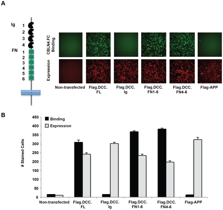
Figure 5. CBLN4 binds to the FN4-6 region of DCC.
A) a schematic representation of DCC is shown with the Ig domains in black and the FN domains in green. Representative fluorescent images are shown for CBLN4-Fc binding to COS-7 cells expressing full-length DCC, DCC Ig1-4, DCC FN1-6, DCC FN4-6, and negative control APP. Detection of CBLN4-Fc binding was through anti-human Alexa Fluor-488 (green). Detection of cell surface expression of all flag-tagged DCC deletion mutants was confirmed with an Alex Fluor-647 labeled anti-mouse IgG (red). B) quantification of CBLN4-Fc binding to the DCC domain deletion constructs. Triplicate independent experiments were performed and error bars represent the standard error of the mean.

