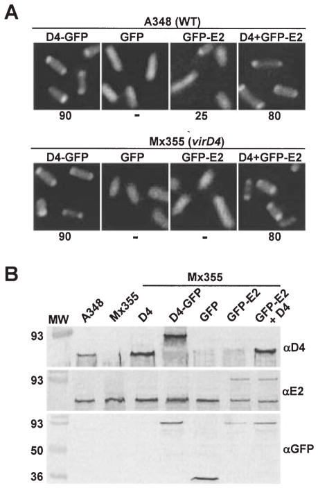
Fig. 1.
VirD4-dependent localization of GFP-VirE2 to A. tumefaciens cell poles.
A. A348 (WT) and Mx355 (virD4 null mutant) cells producing proteins indicated above each panel photographed 10 h after induction with 200 μM AS by fluorescence microscopy. The proteins indicated were synthesized from the following IncP plasmids: D4-GFP (pKA62); GFP (pZDB69); GFP-E2 (pZDB73) and D4 + GFP-E2 (pKA77). The number below each panel represents the percentage of cells with polar fluorescence out of a total of at least 1000 cells examined; the ‘−’ denotes no detectable polar fluorescence.
B. Immunodetection of fusion proteins produced in Mx355 derivatives at 10 h post induction. The proteins listed above each lane were synthesized from the IncP plasmids listed in (A); for D4 (pKA21). Blots were developed with the antisera listed at the right. The reactive species (~60-kDa) in all lanes detected by anti-VirE2 antisera is native VirE2 produced from pTi.

