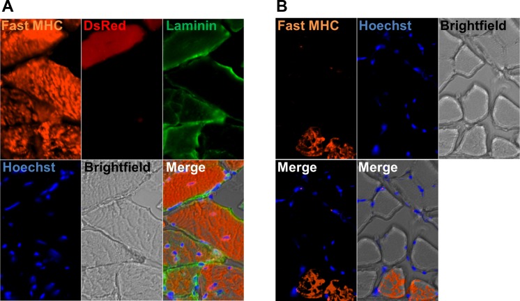
Fig. 8.
DsRed+ muscle fibers in muscles transplanted with DsRed+ type-2 pericytes are fast (type II). A: all fibers express fast myosin heavy chain in TA muscle injected with DsRed+ type-2 pericytes. B: soleus muscle, used as a staining control, also shows some fibers expressing fast myosin heavy chain. A and B show the same area for different channels: Fast MHC staining (orange), DsRed (red), Laminin (green), Hoechst (blue), brightfield, merged fluorescence images, and fluorescence and brightfield merge image; n = 3.

