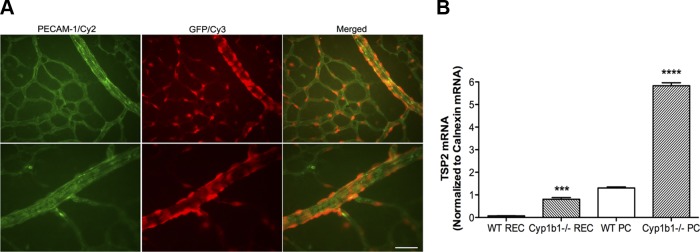
Fig. 1.

Thrombospondin 2 (TSP2) expression is localized to retinal pericytes (PC). A: retinal whole mounts were prepared from P7 TSP2-green fluorescent protein (GFP) transgenic mice and stained with anti-platelet endothelial cell adhesion molecule 1 (PECAM-1) and anti-GFP antibodies. Top and bottom show different fields of view. Scale bar, 20 μm. B: real-time quantitative PCR (qPCR) analysis of relative mRNA expression of TSP2 in wild-type (WT, cyp1b1+/+) and cytochrome P-450 1B1-deficient (cyp1b1−/−) retinal endothelial cells (REC) and PC (n = 9). ***P < 0.001, ****P < 0.0001.
