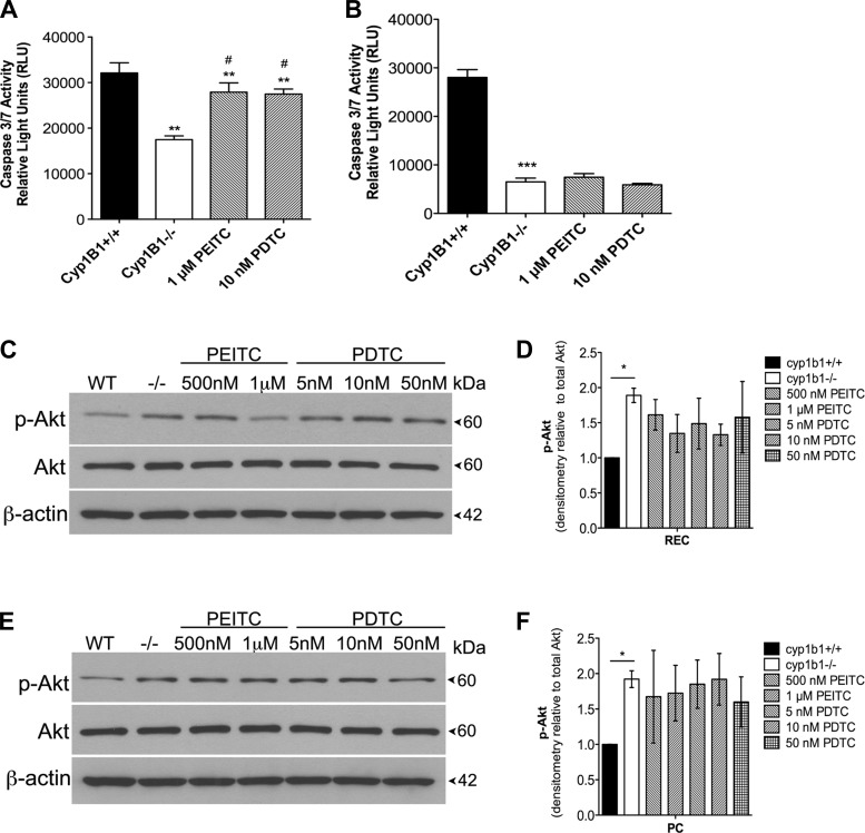
Fig. 3.
Inhibition of NF-κB alters apoptosis in retinal EC. A and B: rate of apoptosis was determined by measuring caspase activity with luminescent signal from caspase-3/7 DEVD-aminoluciferin substrate. In A, cyp1b1−/− EC demonstrated an ∼2-fold decrease in basal levels of caspase-3/7 and a 1.5-fold increase when incubated with PEITC or PDTC. In B, cyp1b1−/− PC demonstrated a 3.5-fold decrease in basal levels of caspase-3/7 and no change when incubated with PEITC or PDTC. **P < 0.01, ***P < 0.001; #P < 0.05 vs. cyp1b1−/− (control). C and E: cyp1b1−/− EC (C) and PC (E) were incubated with PEITC (500 nM and 1 μM) or PDTC (5, 10, and 50 nM), and lysates were analyzed by Western blot analysis for phosphorylated Akt (p-Akt), total Akt, and β-actin. D and F: quantitative assessment of phosphorylated Akt relative to total Akt (n = 2). *P < 0.05.

