Fig. 4.
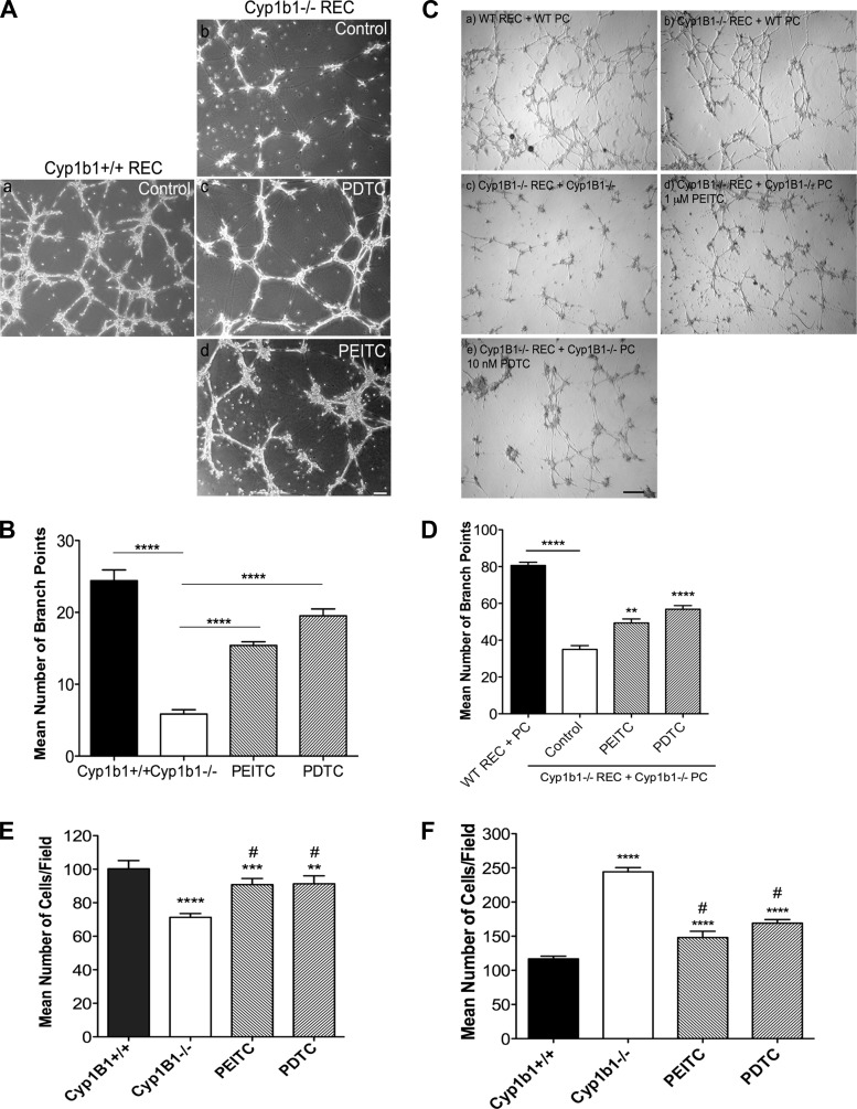
NF-κB suppression restores capillary morphogenesis and migration. A: cyp1b1+/+ and cyp1b1−/− retinal EC were incubated with PEITC and PDTC, plated on Matrigel, and photographed after 18 h. Scale bar, 100 μm. B: mean number of branch points from 5 high-power (×40) fields (n = 3). ****P < 0.0001. C: capillary morphogenesis of retinal EC and PC was assessed by coculturing cells in Matrigel for 18 h. Representative images show cyp1b1+/+ EC + PC (a), cyp1b1−/− EC + cyp1b1+/+ PC (b), cyp1b1−/− EC + cyp1b1−/− PC (c), cyp1b1−/− EC + cyp1b1−/− PC + 1 μM PEITC (d), and cyp1b1−/− EC + cyp1b1−/− PC + 10 nM PDTC (e). Scale bar, 500 μm. D: mean number of branch points (n = 3). **P < 0.01, ****P < 0.0001. E and F: Transwell migration assays after 24 h of incubation of cyp1b1+/+ and cyp1b1−/− EC (E) and PC (F) with PEITC and PDTC (n = 3). Note 30% decrease in migration of cyp1b1−/− compared with cyp1b1+/+ EC, 20% increase in migration of cyp1b1−/− EC incubated with PEITC and PDTC, 75% increase in migration of cyp1b1−/− compared with cyp1b1+/+ PC, 40% decrease in migration of cyp1b1−/− PC incubated with PEITC, and 33% decrease in migration of cyp1b1−/− PC incubated with PDTC. **P < 0.01, ***P < 0.001, ****P < 0.0001; #P < 0.05 vs. cyp1b1−/− (control).

