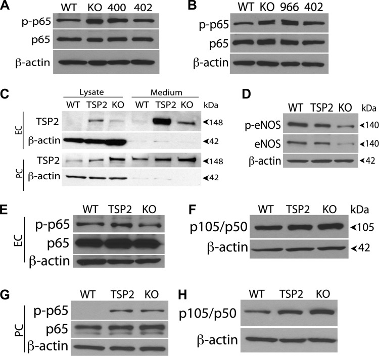
Fig. 8.
TSP2 levels and NF-κB activation are linked. A and B: cyp1b1−/− EC (A) and PC (B) infected with TSP2-specific siRNA were lysed and analyzed for phosphorylated and total p65 by Western blot analysis; β-actin was used as a loading control. Note decreased levels of phosphorylated p65 in cyp1b1−/− [knockout (KO)] EC and PC expressing TSP2-specific siRNA clone 402 with reduced TSP2 level compared with parental cells or those expressing siRNA clone 400 or 966. Also note decreased levels of phosphorylated p65 NF-κB in cyp1b1−/− TSP2 knockdown cells expressing siRNA clone 402 compared with parental control or siRNA clone 400 and 966. C: cyp1b1+/+ EC and PC were transfected using TSP2 expressing adenoviruses. After transfection, conditioned medium and lysates were prepared from EC and PC and analyzed for TSP2 by Western blot analysis. WT, cyp1b1+/+ cells infected with GFP-adenovirus; TSP2, cyp1b1+/+ cells infected with TSP2 adenovirus; KO, cyp1b1−/− cells. D–F: Western blot analysis of phosphorylated endothelial nitric oxide synthase (eNOS), total eNOS, phosphorylated and total p65, and p105/p50 in cyp1b1+/+ EC. Note reduced level of eNOS and phosphorylated eNOS in cyp1b1+/+ cells expressing TSP2 compared with control cells. G and H: Western blot analysis of phosphorylated and total p65 and p105/p50 in cyp1b1+/+ PC expressing TSP2. Note increased phosphorylated p65 NF-κB in cyp1b1+/+ PC expressing TSP2 compared with control.

