
Figure 1.
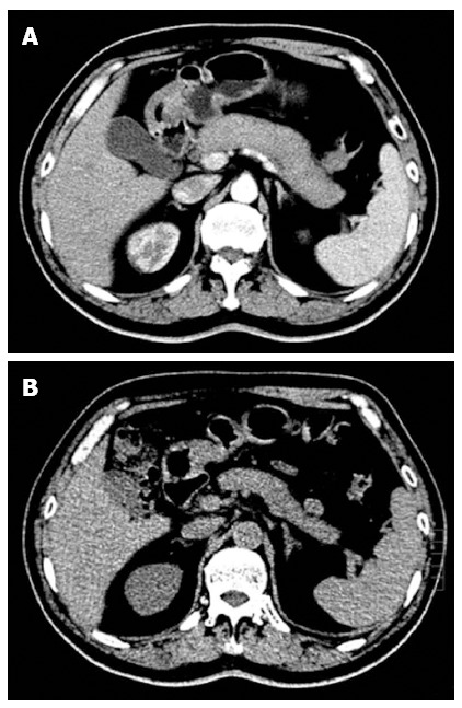
Typical imaging features of type 1 autoimmune pancreatitis. Computed tomography (CT) scan showing diffuse swelling of the pancreas with loss of lobulation (A), and a dramatic decrease in swelling of the pancreas after 3 wk of steroid treatment (B).