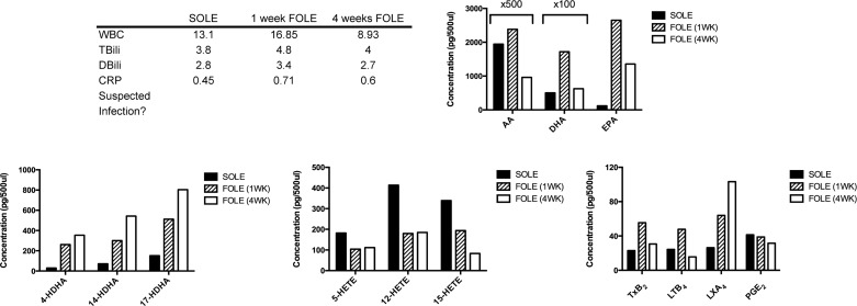
Fig. 6.
Bioactive LM profile for patient 4. Table outlines selected laboratory values collected immediately prior to transition from SOLE to FOLE, 1 wk after FOLE, and 4 wk after FOLE and notes whether infection was suspected at the time of sample collection. See Fig. 3 legend for abbreviations and units of measure. Bar graphs display results from analysis of PUFAs, Rv precursors (HDHA), HETEs, and eicosanoids. Columns are labeled with multiplicative values to indicate changes in scale appropriate for the column and denote a concentration equal to the displayed value multiplied by the factor. Rv precursor compounds (4-, 14-, and 17-HDHA) rose progressively with greater duration during FOLE administration. Proresolving mediator LXA4 increased with FOLE administration.

