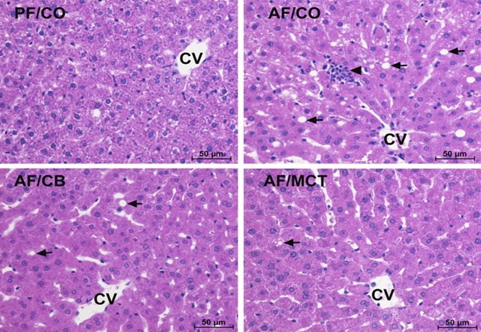
Fig. 1.
Liver histopathology in rats fed ethanol with different dietary fats for 8 wk. Liver sections were stained by hematoxylin and eosin. Light microscopy showed lipid accumulation (arrows) and inflammatory cell infiltration (arrowheads) in the liver. Scale bar: 50 μm. CV, central vein; PF/CO, pair fed with corn oil; AF/CO, alcohol fed with corn oil; AF/CB, alcohol fed with cocoa butter; AF/MCT, alcohol fed with medium-chain triglycerides.

