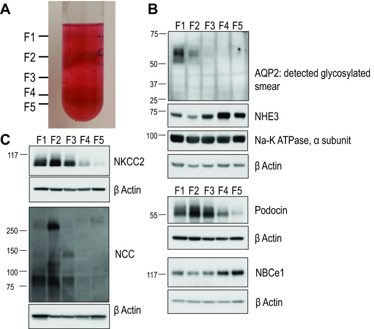
Fig. 1.
Characteristics of our renal cortical tubule preparation. A: a mixture of renal tubules from the rabbit kidney cortex was applied to a 45% self-forming Percoll gradient and subjected to centrifugation at 25,000 × g for 35 min at 4°C. Tubules typically migrated into tubule fractions F1–F5, with proximal tubules (PTs) migrating in lower tubule fractions F4 and F5. B and C: characterization of tubule fractions F1–F5 by Western blot. Lysates were prepared from material isolated from tubule fractions F1–F5, resolved by SDS-PAGE, transferred to polyvinylidene difluoride (PVDF) membranes, and probed with the indicated antibodies. From top to bottom in B: aquaporin 2 (AQP2; collecting duct marker), Na+/H+ exchanger 3 (NHE3; brush-border membrane), α-subunit of Na+-K+-ATPase (basolateral transporter, renal tubule epithelium), β-actin (loading control), podocin (podocyte marker), β-actin (loading control), Na+-HCO3− cotransporter (NBC)e1-A (PT marker), and β-actin (loading control). From top to bottom in C: Na+-K+-Cl− cotransporter 2 (NKCC2; thick ascending limb marker), β-actin (loading control), Na+-Cl− cotransporter (NCC; distal convoluted tubule marker), and β-actin (loading control).

