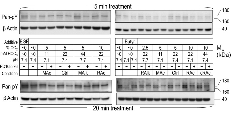
Fig. 3.
Pan-pY immunoreactivity at 180 and 160 kDa in rabbit PT preparations subjected to acid-base disturbances (5 and 20 min). Tubules were exposed to one of several treatment solutions, and protein was subjected to Western blot analysis. For each lane, we show (from top to bottom) the pan-pY immunoreactivity from tubules that were exposed to treatments for 5 min and a reprobe of the same blot with an anti-actin antibody (top), a summary of the treatment conditions (table in middle), and the pan-pY immunoreactivity from tubules that were exposed to treatments for 20 min and a reprobe of the same blot with an anti-actin antibody (bottom). MAc, metabolic acidosis; MAlk, metabolic alkalosis; RAc, respiratory acidosis; RAlk, respiratory alkalosis; cRAc, fully compensated respiratory acidosis.

