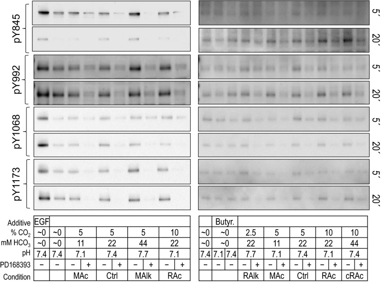
Fig. 6.
ErbB1 immunoreactivity at individual pY sites in rabbit PT preparations subjected to acid-base disturbances (5 and 20 min). Tubules were exposed to one of several treatment solutions, and protein was subjected to Western blot analysis. For each lane, we show (from top to bottom) pY845 at 5 and 20 min, pY992 at 5 and 20 min, pY1068 at 5 and 20 min, pY1173 at 5 and 20 min, and a summary of the treatment conditions. Note that the blots on the right repeat some of the conditions on the left. We do not show the reprobes of the blots for actin.

