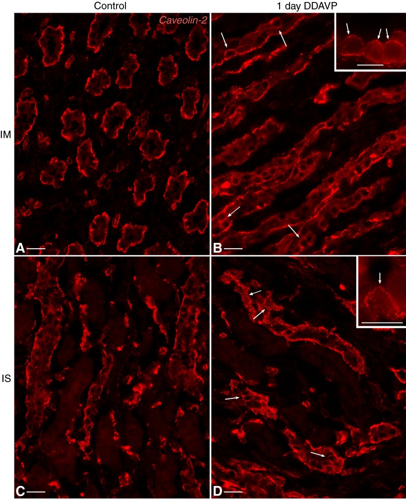
Fig. 10.
Distribution of Cav2 in PCs of the BB rat renal CD. Cav2 (red) localization in IMCD (A) and ISCD (C) PCs was mainly basolateral, similar to that of Cav1. After 24 h of DDAVP treatment, Cav2 appeared in the apical membrane domain of certain PCs (arrows) from both the IMCD (B) and ISCD (D) as a number of small, scattered spots. Insets show higher-magnification images of cells, demonstrating examples of apical Cav2 staining (arrows). Bars = 20 μm; bars in insets = 10 μm.

