Fig. 3.
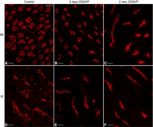
Subcellular localization of aquaporin-2 (AQP2; red) in control and DDAVP-treated BB rats. AQP2 had a diffuse localization in inner medullary CD (IMCD) PCs of control BB rats (A), which became polarized to the apical membrane of these cells after treatment with DDAVP for 3 days (B) or 2 wk (C). Similarly, ISCD PCs exhibited cytosolic diffuse AQP2 in control animals (D) but sharp apical AQP2 localization after 3 days (E) or 2 wk (F) of DDAVP treatment. Bars = 25 μm.

