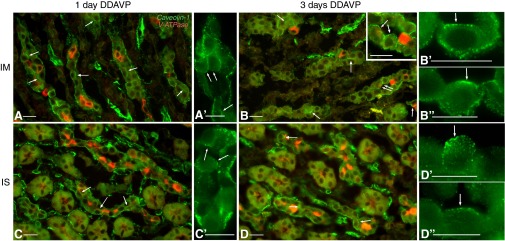
Fig. 4.

Double labeling for Cav1 (green) and V-ATPase (red) in the kidney of BB rats after short-term DDAVP treatment. After 1 day of DDAVP exposure, Cav1 was predominantly located in the basolateral membrane domain of PCs (identified by their negative labeling for V-ATPase) in both the IMCD (A and A′) and ISCD (C and C′). At this time point, spots of apical Cav1 staining started appearing in a few cells (arrows). After 3 days, PCs with significant apical staining for Cav1 were readily found in both the IMCD (B) and ISCD (D). The inset in B shows a higher-magnification image of the region indicated by the double arrow, emphasizing apical staining (arrows) facing the open lumen of the tubule. In even higher-magnification panels (B′ and B″ for the IMCD; D′ and D″ for the ISCD), PCs in open tubules from BB rats treated with DDAVP for 3 days showed apical Cav1 staining (arrows). Bars = 15 μm.
