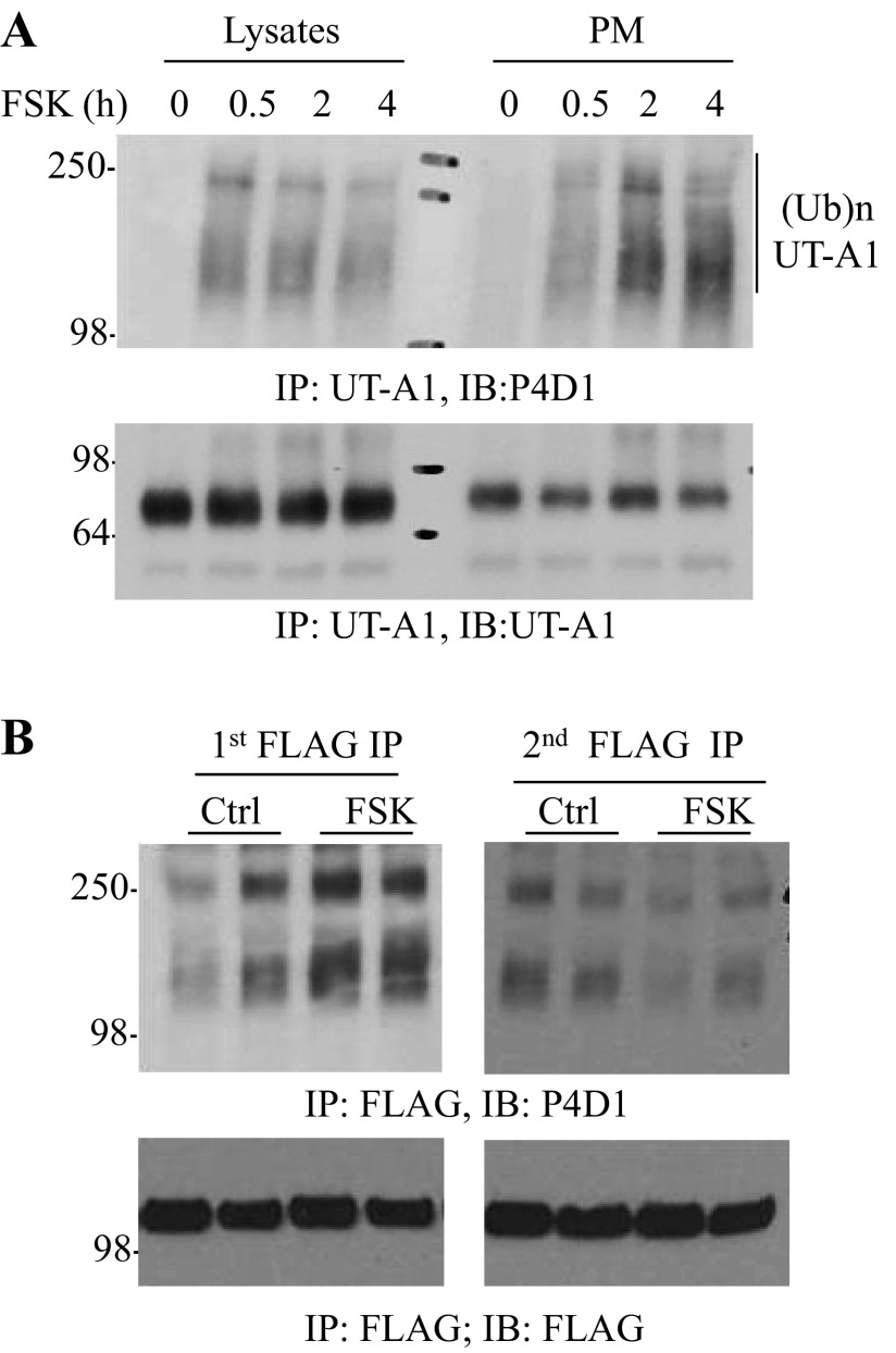
Fig. 2.
FSK stimulation promotes cell surface UT-A1 ubiquitination. A: UT-A1 MDCK cells were treated with 10 μM FSK for different times. The plasma membrane (PM) was isolated by a sucrose gradient ultracentrifugation. Equal amounts of lysates or the PM were immunoprecipitated with UT-A1 antibody followed by immunoblotting with antiubiquitin (Ub) antibody P4D1. B: FLAG-Tac-UT-A1 transfected HEK 293 cells were treated without or with 10 μM FSK for 4 h at 37°C. The cells were incubated with FLAG antibody on ice for 1 h then lysed in RIPA buffer. The lysates were incubated with protein G beads overnight (first immunoprecipitation of the cell surface UT-A1). The supernatant was collected and processed for a second immunoprecipitation with FLAG antibody (cytoplasmic UT-A1). The immunoprecipitated samples were analyzed by Western blot with ubiquitin antibody. This experiment was repeated two times with similar results.

