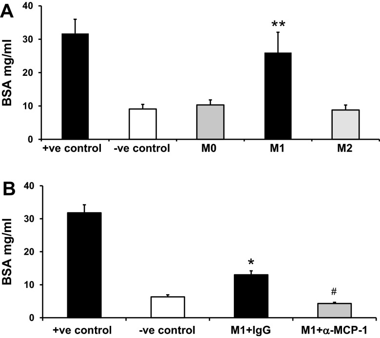
Fig. 8.
M1 macrophages increased podocyte permeability in vitro. A and B: nouse bone marrow monocytes were isolated from C57BL6/J mice and cultured in RPMI-1640 supplemented with 10 ng/ml macrophage colony-stimulating factor for 7 days in six-well plates followed by M1 and M2 macrophage induction. Differentiated podocytes were cultured in Transwell membranes. Macrophages and podocytes were cocultured in the Transwell system to study the transepithelial passage of BSA during a 6-h period. Upper chamber media were collected to measure BSA concentration. The upper chamber membrane without podocytes was used as the positive control; no macrophages in the lower chamber was used as the negative control. A: macrophages without stimulation were defined as M0. B: M1 macrophages were seeded with IgG or anti-mouse MCP-1. Results are means ± SE. *P < 0.05 and **P < 0.001 compared with the negative control; #P < 0.01 compared with M1 macrophage + IgG.

