
Figure 2.
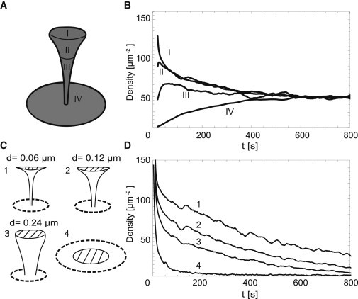
Spine shape influences exit dynamics of receptors. (A) Domains used to analyze density evolution in panel B (R = 1 μm, B = 4 μm, d = 0.12 μm, D = 0.1 μm2/s; see Fig. 1C). (B) Time evolution of receptor density in domains I–IV after the release of 1000 receptors at the top of the spine. Reflecting boundary was positioned at a distance of 1 μm from the center of the base of the spine. (C) Four different spine geometries used in panel D. (1–3: R = 1 μm, B = 4 μm, D = 0.1 μm2/s; 4: RPSD = 0.71 μm, R1 = 2.35 μm, D = 0.1 μm2/s; see Fig. 1C.) (D) Time evolution of receptor density (dashed area). One-thousand receptors were released in the center of this region at t = 0.