
Figure 4.
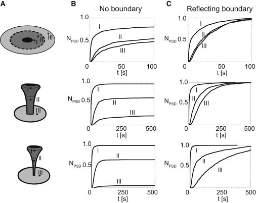
Position of exocytosis and shape of the spine strongly alter the dynamics and efficiency of receptor capturing by the PSD. (A) Three different geometries used to analyze effect of the exocytotic position (R = 1 μm, B = 4 μm, D = 0.1 μm2/s; see Fig. 1C). (B and C) Fraction of absorbed receptors over time for three different shapes after exocytosis at positions I, II, or III, as depicted in panel A. Reflecting boundaries were positioned at infinity (B) or at a distance of 1 μm from the center of the base of the spine (C). One-thousand receptors are released on various positions (close to the head of the spine (I), in the neck of the spine (II), and on a 0.5-μm distance from the centrum of the base of the spine on the dendritic membrane (III)). We consider three different shapes, these being a spine with a neck radius of 0.06 μm, 0.24 μm, and a planar shape. In the flat geometry the total surface area in the PSD/Spine/Dendrite is the same as in the spine, with a neck radius of 0.24 μm (release I at 1.2 μm, II at 2.2 μm, and III at 2.4 μm from the center). These figures show that, especially for mushroomlike spines, exocytosis close to the top of the spine results in a much faster and more efficient capturing of receptors. To see this figure in color, go online.