
Figure 1.
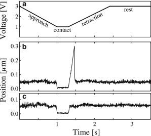
Typical probe bead tracking curves. (a) Piezo control motion cycle. (b) Probe bead tracking in case of a binding-rupture event. As piezo voltage decreases, the test bead is brought into contact with the probe bead, and the motion is stopped for 0.3 s to allow for bond formation. During contact, the erythrocyte is slightly compressed, and the probe bead is pushed away from its rest position (impingement force 6 pN). During retraction, the probe bead is pulled by the test bead until the point of bond rupture (force loading rate 286 pN/s, rupture force 39 pN). Finally, the probe bead returns to its rest position. (c) Probe bead tracking in the case of no bond formation, impingement force 8 pN. Probe stiffness was 150 pN/μm in panels b and c.