
Figure 1.
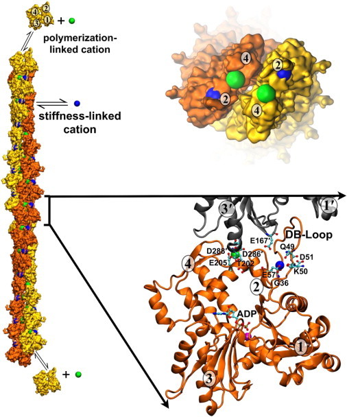
Location of two discrete, actin filament-specific cation-binding sites. The actin filament structure is based on the model of Fujii et al. (50) and includes the predicted cation-binding sites from Kang et al. (18). The barbed end of the filament is toward the bottom of the figure and the pointed end with associated cations is shown following a 90° rotation. (Yellow and orange) Surface rendering of actin monomers; (orange and gray) cartoon rendering of actin monomers. (Numbers in ovals) Actin monomer subdomains. (Green) Polymerization site cation; (blue) stiffness site cation; (magenta) nucleotide-associated cation. (Ball-and-stick representation) ADP nucleotide and specific cation-binding residues. To see this figure in color, go online.