Fig. 3.
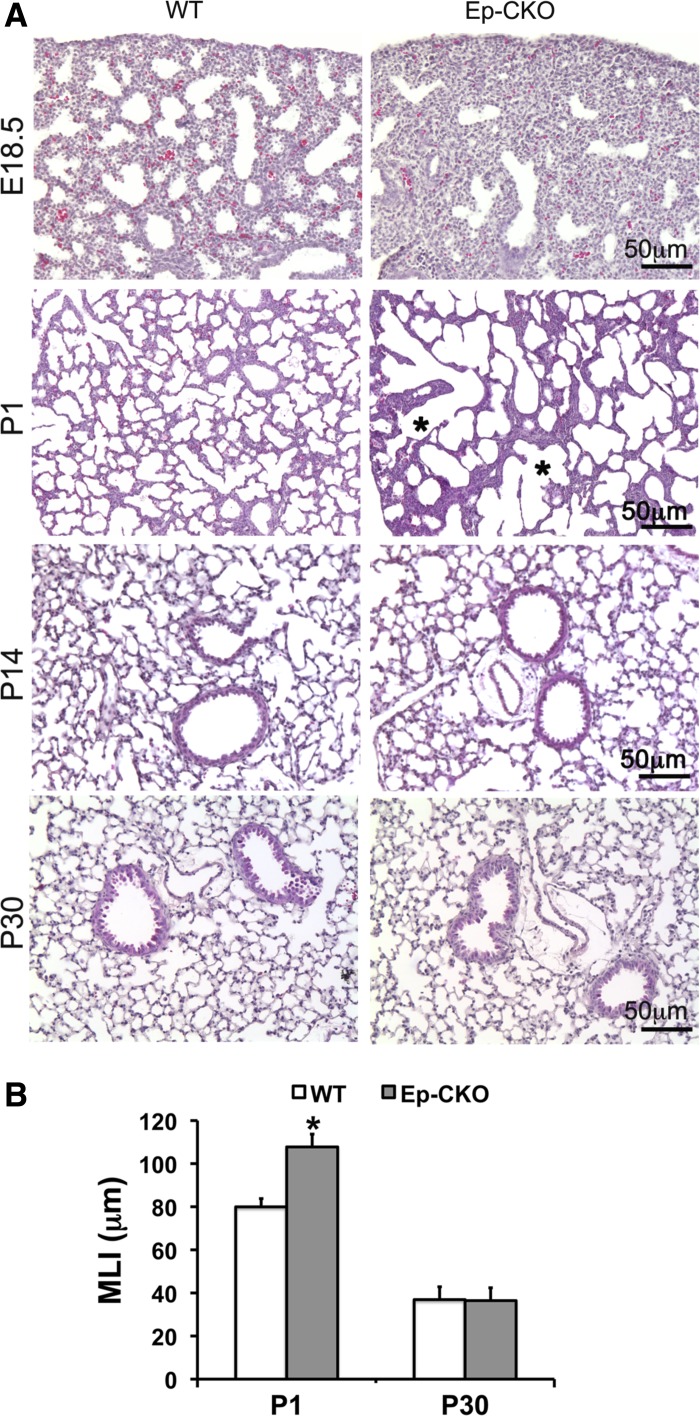
Lung epithelial-specific deletion of TACE resulted in abnormal perinatal lung development. A: hematoxylin and eosin (H&E)-stained lung tissue section of TACE Ep-CKO mice and WT littermates at different developmental stages. The diameters (shown by asterisk) of pulmonary alveoli in P1 TACE Ep-CKO lung were significantly increased compared with WT controls. B: morphometric analysis of TACE Ep-CKO lung by mean linear intercept (MLI) at different development stages. The MLI was significantly larger in P1 TACE Ep-CKO lungs (107.77 ± 5.95 μm) than that of wild-type controls (79.95 ± 3.84 μm, P < 0.01). After postnatal lung alveolarization (P14 and P30), the morphological difference between TACE Ep-CKO and wild-type control was no longer detected (MLI: 36.44 ± 0.91 μm in TACE Ep-CKO lung vs. 36.89 ± 0.17 μm in WT control at P30). *P < 0.05 (n = 5 for each genotype at the specified age).

