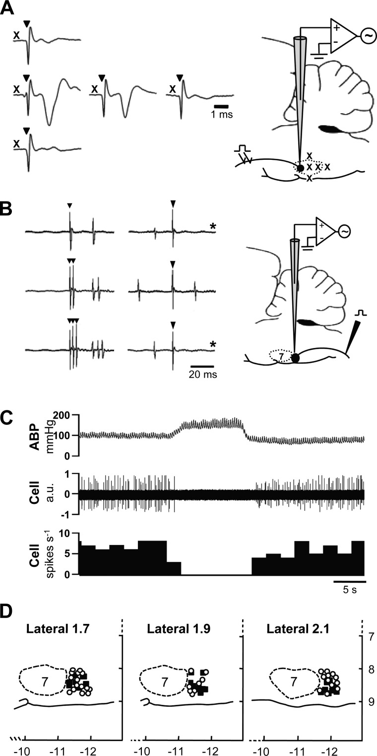
Fig. 2.
Identification and distribution of rostral ventrolateral medulla (RVLM) neurons. A: RVLM vasomotor neurons are clustered just caudal to the facial nucleus, the caudal pole of which was mapped according to the amplitude of the antidromic field potential evoked by electrical stimulation of the facial nerve (CN VII). X, approximate location of recording sites (see brain stem profile to the right); ▼, stimulus artifacts. Note that the amplitude of the antidromic field potential decays as the recording electrode is moved to more posterior sites within the facial nucleus and disappears when the most caudal boundary of the nucleus is reached. B: RVLM vasomotor neurons have axonal projections to spinal cord. Spinally projecting neurons (NT, n = 9; HT, n = 6) were identified by recording antidromic spikes evoked by stimulation of the T2 spinal segment. The antidromic nature of evoked spikes was confirmed by the consistent onset latency of spikes relative to each stimulus artifact (▼, at left and right), their ability to follow high frequency (333 Hz) stimulation (middle and lower traces at left), and collision with spontaneous action potentials (at right; *sweeps where collision occurred). C: spontaneous discharge of recorded RVLM neurons was consistently inhibited by raising arterial blood pressure (ABP). D: recorded RVLM neurons in NT (○) and HT (■) rats were similarly distributed and consistently located within ∼700 μm of the caudal pole of the facial nucleus, 1.7–2.1 mm lateral to midline and within 1.0 mm of the ventral surface of brain. Note that x- and y-axis values are distances (in mm) relative to bregma and brain surface, respectively. AU, arbitrary units.

