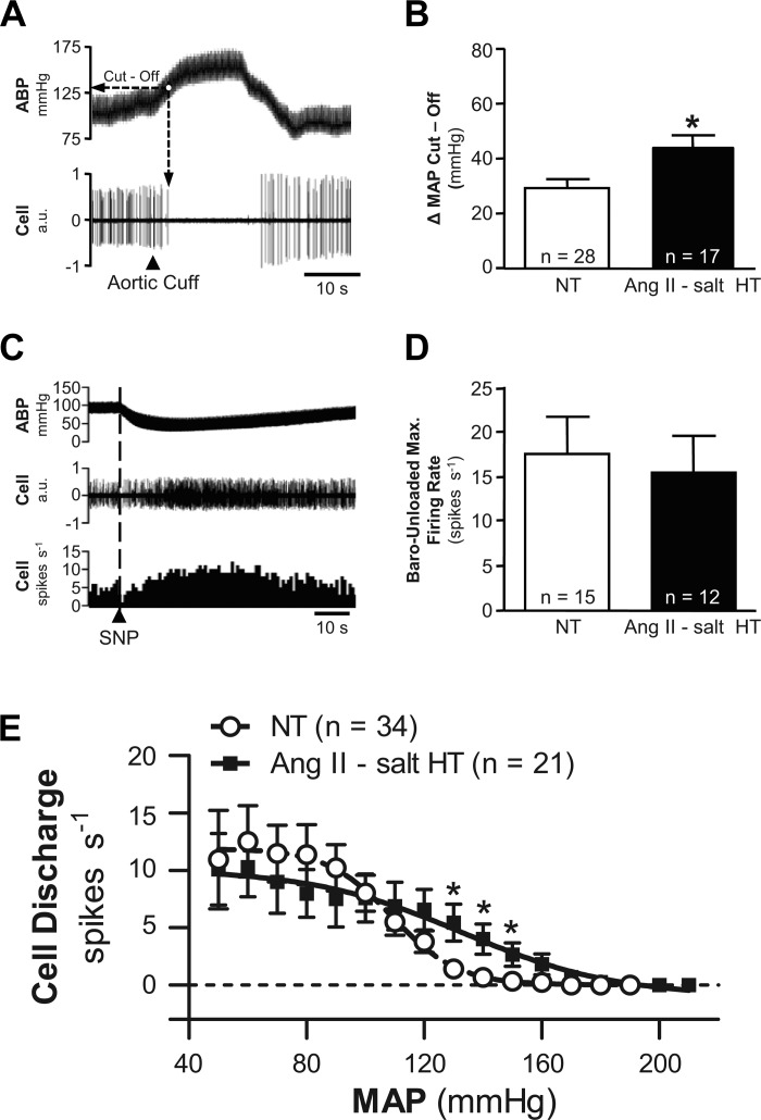
Fig. 4.
Comparison of barosensitivity of RVLM neuronal discharge. A: example data show that raising ABP by inflation of an aortic cuff led to silencing of discharge. The change of MAP required to silence cell discharge was deemed “ΔMAP Cut-Off.” B: summary data for 28 neurons from NT rats (white bar) and 17 neurons from HT rats (black bar) indicate that a significantly (*P < 0.01) greater increase of MAP was required to fully silence neurons in the HT group. C: example traces show that intravenous injection of the vasodilator sodium nitroprusside (SNP) consistently lowered AB and increased discharge of identified RVLM neurons. D: summary data of maximum firing rate achieved during SNP-induced unloading of arterial baroreceptors. Note that discharge increased similarly among neurons in NT (white bar) and HT (black bar) rats. E: summary baroreflex function curves relating MAP to RVLM discharge frequency. Note that NT (○) and HT (■) groups had similar maximum discharge frequency during baroreceptor unloading and similar minimum discharge frequency when MAP as raised. Neurons in the ANG II-salt HT group showed a significant degree of resistance to baroinhibition across values of MAP ranging from 120 to 150 mmHg.

