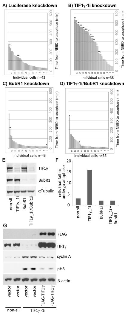
Figure 7. Inactivation of the SAC allows TIF1γ knockdown cells to pass through mitosis from NEBD to anaphase.
HeLa cells expressing Histone H2B mCherry and α-tubulin-EGFP were treated with either an siRNA specific for luciferase (A) or siRNAs specific for TIF1γ (B), BubR1 (C) or TIF1γ and BubR1 (D). 48h post-knockdown cells were subjected to a thymidine block for 16 h, whereupon cells were released back into cycle and video imaging began 9 h later. Bar charts depicting time taken to pass from NEBD to anaphase are presented. * represents cells that failed to undergo successful metaphase-to-anaphase transition during filming (depicted graphically in Fig. 7F). (E) Western blots for TIF1γ BubR1 and α-tubulin in cells treated with luciferase, TIF1γ BubR1, or γ and BubR1 siRNAs. (G) Expression of a siRNA-resisitant TIF1γ species allows for cyclin A degradation and mitotic progression. HeLa cells were treated with either non-silencing siRNA, or siRNAs specific for TIF1γ for 48 h. Cells were then transfected with vector alone or a construct expressing FLAG-TIF1γ and cultured for a further 24h. Cell lysates were prepared, and the levels of FLAG-TIF1γ, endogenous TIF1γ, cyclin A, phospho Ser10-histone H3 (pH3) and β-actin were all determined by Western blot.

