
Fig 1.
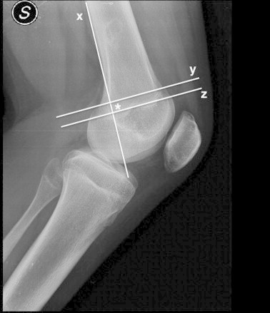
The method of Schöttle et al.3 used to obtain the anatomic femoral MPFL attachment, as shown on a radiograph in a left knee. A straight lateral view of the knee is obtained intraoperatively with an image intensifier. The radiographic landmark of the femoral attachment is determined by 3 lines: an elongation of the posterior femoral cortex (line x) and 2 lines perpendicular to line x, 1 passing the origin of the medial condyle (line y) and 1 passing the most posterior aspect of the Blumensaat line (line z). The landmark is anterior to line x and between lines y and z (asterisk).