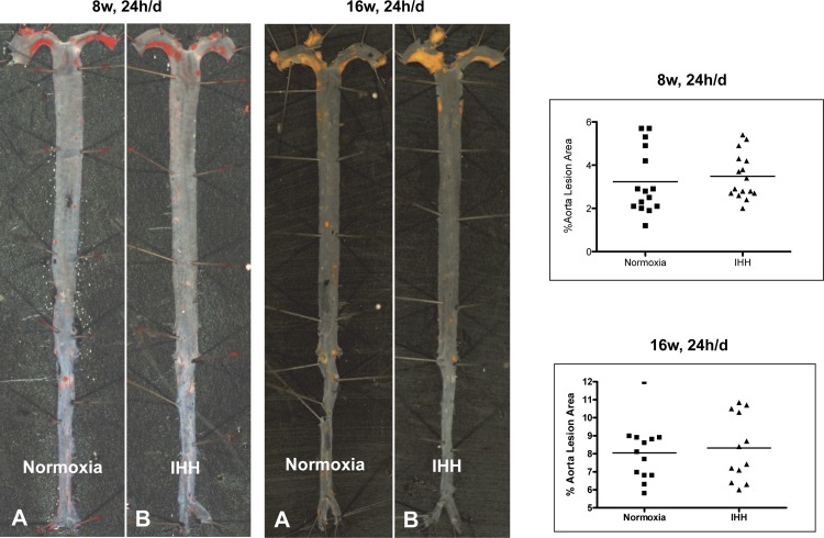
Fig. 1.
Lesion development in Normoxia and intermittent hypoxia/hypercapnia (IHH) aortas. Mice were exposed to IHH for indicated times and en face atherosclerosis quantified as explained in methods. Representative aortas from mice exposed to IHH for 24 h/day for 8 wk and 16 wk are shown, as well as the values for all mice in these experimental protocols. Formation of lesions in control and IHH-induced mice: Ldlr−/− mice that were exposed to WD and either kept in room air (normoxia) or exposed to IHH for 8 wk and 16 wk. Sudan IV-stained aortas showed no difference in lesion area. Data are presented as mean ± SD.

