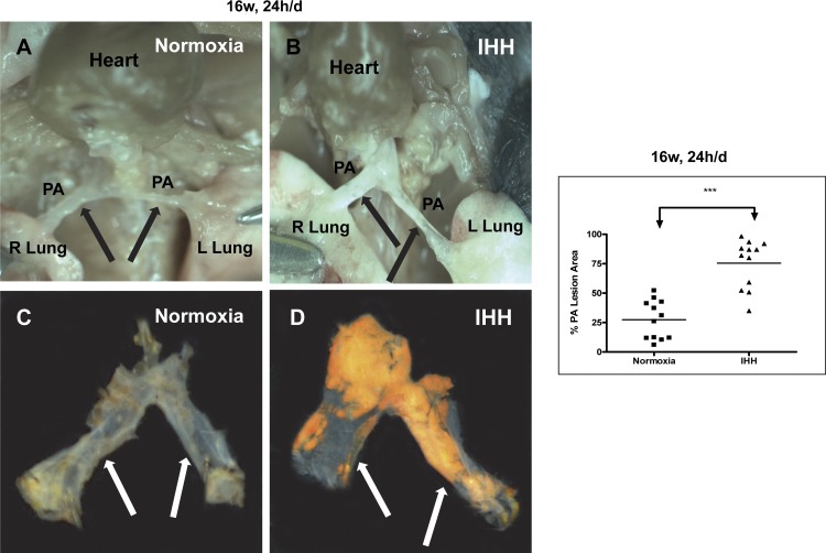
Fig. 4.
Lesion development in the pulmonary arteries of Normoxia and IHH mice exposed to IHH for 16 wk, 24 h/day. A and B show photographs of the in situ appearance of pulmonary arteries (PAs) of a Normoxia (A) and IHH (B) mouse, while C and D show the appearance of the arteries after dissection and Sudan IV staining. The graph shows the percent of the PA stained by Sudan IV for each of the mice examined. ***P < 0.0001. Data are presented as means ± SD.

