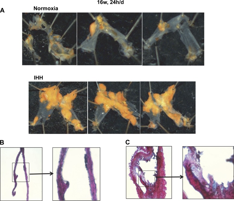
Fig. 5.
Additional examples of pulmonary arteries dissected from Normoxia mice (A) and IHH mice (B) after 16 wk, 24 h/day exposure. Pulmonary arteries were stained with Sudan IV. B and C display photomicrographs of modified Gieson stained paraffin sections of PA from a Normoxia (B) or IHH (C) mouse (433 magnification), along with a blowup of the indicated portion of each artery section.

