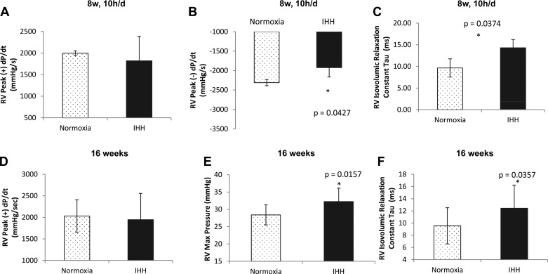
Fig. 7.
Hemodynamic properties of the right ventricle in mice exposed for 8 wk, 10 h/day and for 16 wk. Hemodynamic analysis of the right ventricle in mice exposed to IHH for 8 wk, 10 h/day revealed a decrease in peak (−) dP/dt (B), and an increase in the isovolumic relaxation constant, tau (C) while there was no difference in peak (+) dP/dt (A). In mice exposed to IHH for 16 wk, there was an increase in RV max pressure (E) and an increase the isovolumic relaxation constant, tau (F), while there was no difference in peak (+) dP/dt (D). Data are presented as means ± SD.

